Histology Coloring Book Navigation
  • Home
  • Chapters
  • About Project
  • Our Team
    • Project Leads
    • Partner Project Lead
    • Illustrator
    • Reviewers
    • Design Contributors
  • Contact Us
  • Terms of use
  • Search
  • Home
  • Chapters
  • About Project
  • Our Team
    • Project Leads
    • Partner Project Lead
    • Illustrator
    • Reviewers
    • Design Contributors
  • Contact Us
  • Terms of use
  • Search

Trachea & Respiratory Epithelium

  • Respiratory System
Image
Coloring Image
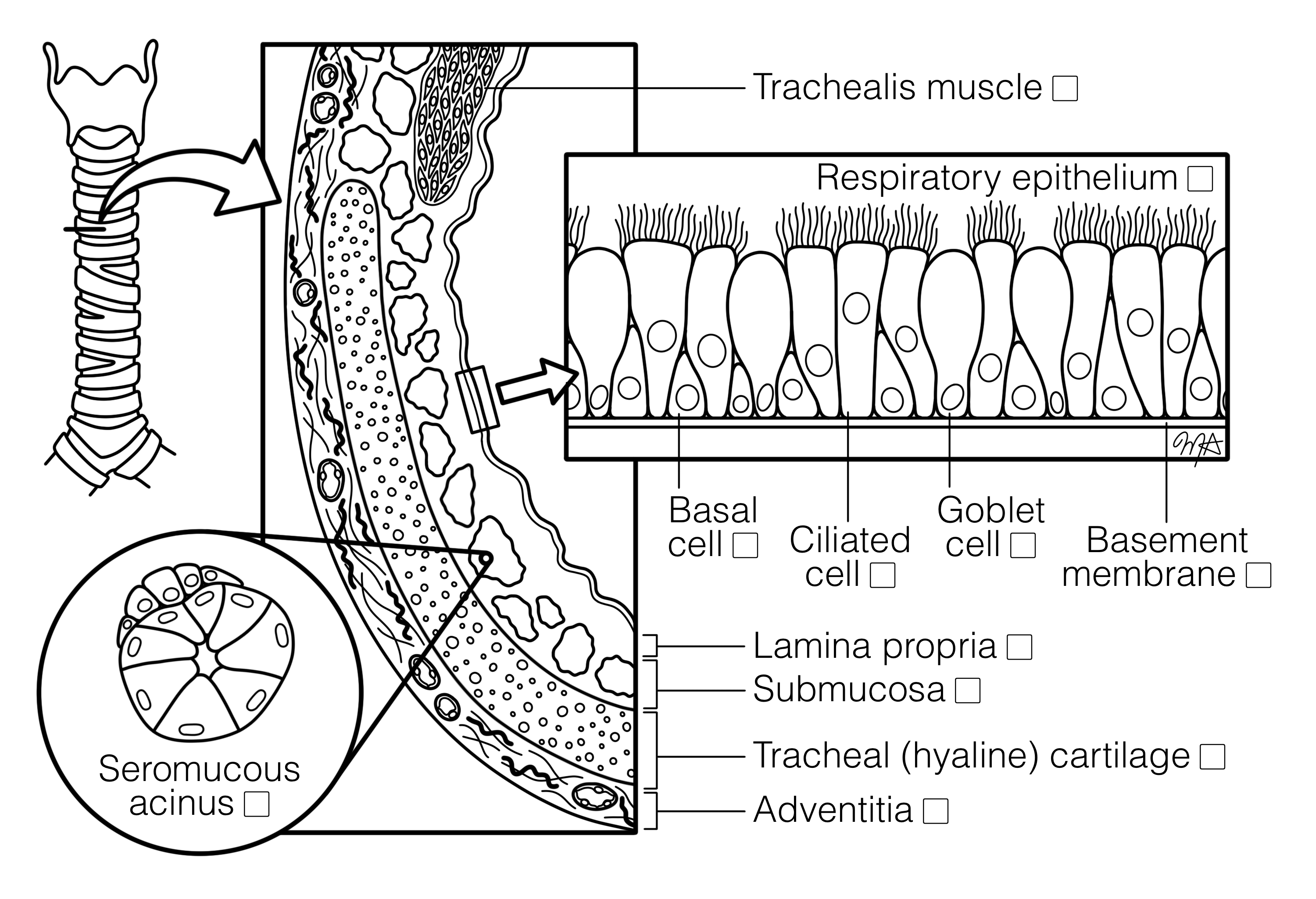
Histological section of the trachea stained with H&E, showing the layered structure of the airway wall. The mucosa is lined by pseudostratified ciliated columnar epithelium with goblet cells, basal cells, and a distinct basement membrane, highlighted in an inset. Beneath the lamina propria lies the submucosa containing seromucous glands, shown in a separate inset. The tracheal wall includes hyaline cartilage rings and the trachealis muscle posteriorly, with adventitia forming the outer layer. The image illustrates the organization of the trachea and its specialized respiratory epithelium.
Histology Image
University of Michigan, M. Hortsch


    Download Coloring Image
    Download Histology image

    All Chapters


    • Blood Cells
      5

    • Cardiovascular System
      5

    • Cells and Organelles
      2

    • Connective Tissues
      3

    • Digestive and Accessory Organs
      12

    • Endocrine System
      3

    • Epithelial Tissues
      4

    • Immune System
      6

    • Integumentary System
      1

    • Muscle Tissues
      8

    • Nervous Tissue
      10

    • Reproductive Systems
      13

    • Respiratory System
      4

    • Skeletal and Articular Systems
      10

    • Special Senses
      10

    • Urinary System
      9
    X
    • About Project
    • Reviewers
    • Design Contributors
    • Illustrator
    • Project Leads
    • Contact Us
    • Table Of Content
    • Glossary
    • Admin

    DESIGNED AND DEVELOPED BY ENGR DIGITAL