Histology Coloring Book Navigation
  • Home
  • Chapters
  • About Project
  • Our Team
    • Project Leads
    • Partner Project Lead
    • Illustrator
    • Reviewers
    • Design Contributors
  • Contact Us
  • Terms of use
  • Search
  • Home
  • Chapters
  • About Project
  • Our Team
    • Project Leads
    • Partner Project Lead
    • Illustrator
    • Reviewers
    • Design Contributors
  • Contact Us
  • Terms of use
  • Search

Terminal Respiratory Unit

  • Respiratory System
  • Alveoli
Image
Coloring Image
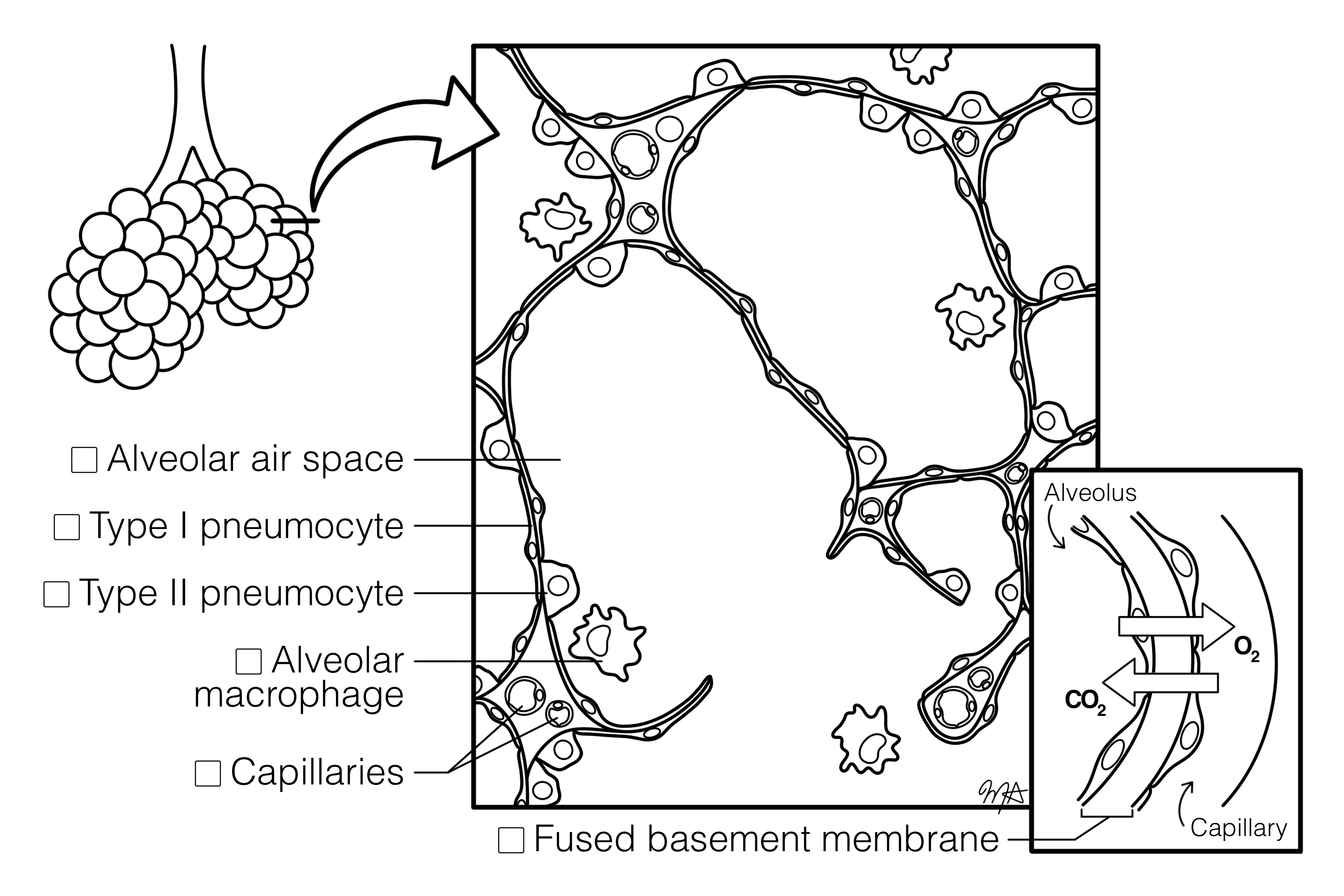
Histological section of the lung stained with H&E, showing the terminal respiratory unit. Alveolar air spaces are separated by thin septa containing capillaries. Type I pneumocytes form most of the alveolar lining, while Type II pneumocytes with rounded nuclei are interspersed and responsible for surfactant secretion. Alveolar macrophages (dust cells) are present within the air spaces. The respiratory membrane (air-blood barrier) is visible where capillary endothelium and alveolar epithelium meet. The image illustrates the structural components of the alveolus and the gas-exchange interface.
Histology Image
University of Tasmania, J. Chapman


    Download Coloring Image
    Download Histology image

    All Chapters


    • Blood Cells
      5

    • Cardiovascular System
      5

    • Cells and Organelles
      2

    • Connective Tissues
      3

    • Digestive and Accessory Organs
      12

    • Endocrine System
      3

    • Epithelial Tissues
      4

    • Immune System
      6

    • Integumentary System
      1

    • Muscle Tissues
      8

    • Nervous Tissue
      10

    • Reproductive Systems
      13

    • Respiratory System
      4

    • Skeletal and Articular Systems
      10

    • Special Senses
      10

    • Urinary System
      9
    X
    • About Project
    • Reviewers
    • Design Contributors
    • Illustrator
    • Project Leads
    • Contact Us
    • Table Of Content
    • Glossary
    • Admin

    DESIGNED AND DEVELOPED BY ENGR DIGITAL