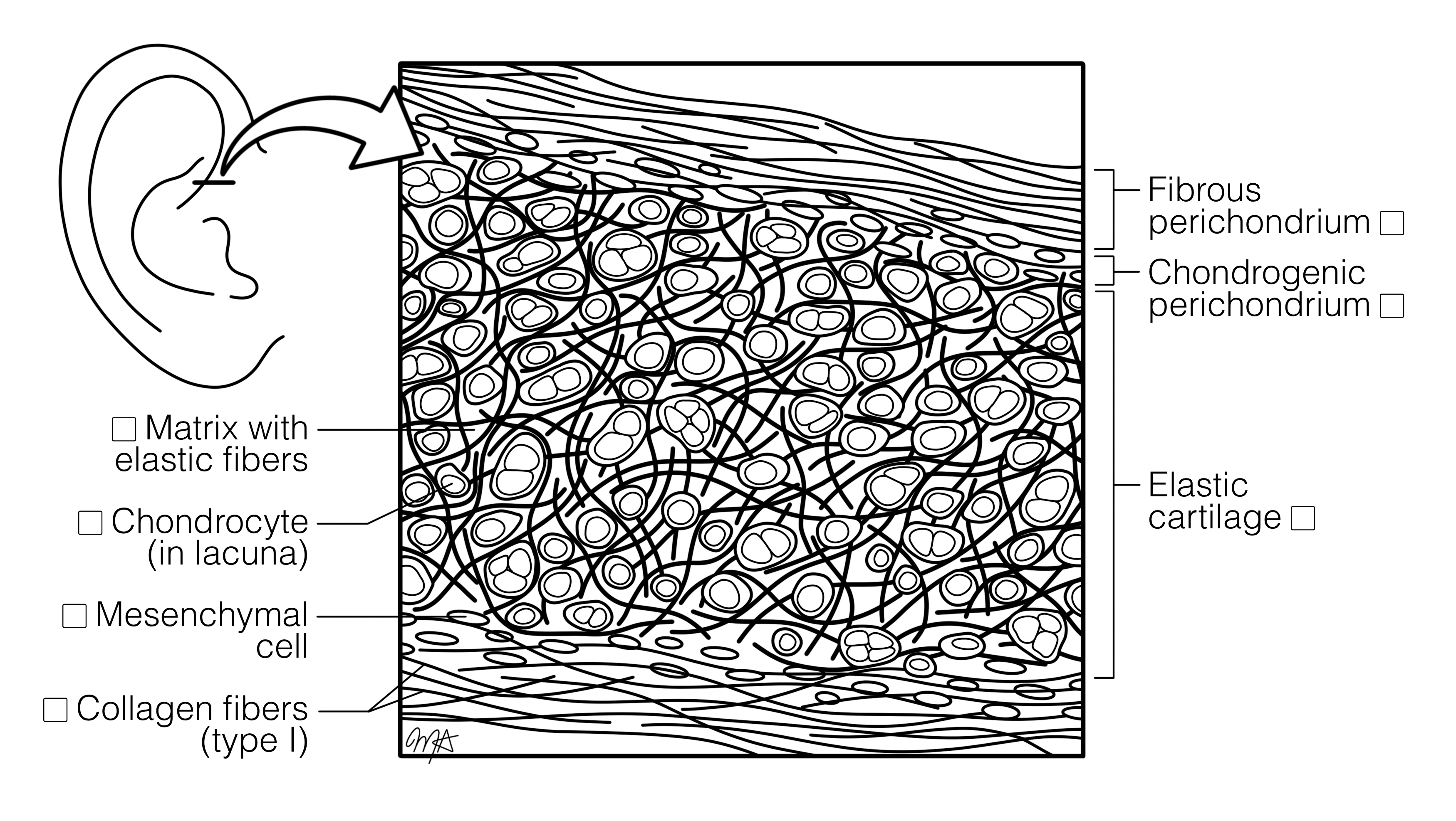
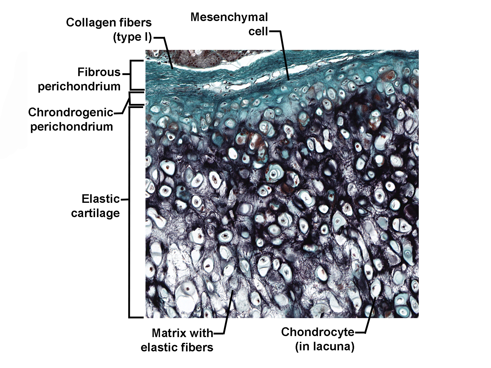
Histology Coloring Book Navigation
  • Home
  • Chapters
  • About Project
  • Our Team
    • Project Leads
    • Partner Project Lead
    • Illustrator
    • Reviewers
    • Design Contributors
  • Contact Us
  • Terms of use
  • Search
  • Home
  • Chapters
  • About Project
  • Our Team
    • Project Leads
    • Partner Project Lead
    • Illustrator
    • Reviewers
    • Design Contributors
  • Contact Us
  • Terms of use
  • Search

Elastic Cartilage (Ear/Auditory Tube)

  • Skeletal and Articular Systems
  • Cartilage Tissue
Image
Coloring Image
Histological section of elastic cartilage stained with a special connective tissue stain, showing chondrocytes within lacunae embedded in a matrix rich in elastic fibers. The cartilage is surrounded by a perichondrium with two layers: an outer fibrous layer containing type I collagen fibers and mesenchymal cells, and an inner chondrogenic layer adjacent to the cartilage. The elastic cartilage matrix appears dark due to elastic fibers, and chondrocytes are distributed throughout. The image illustrates the structural organization of elastic cartilage and its perichondrium.
Histology Image
University of British Columbia, K. Pinder


    Download Coloring Image
    Download Histology image

    All Chapters


    • Blood Cells
      5

    • Cardiovascular System
      5

    • Cells and Organelles
      2

    • Connective Tissues
      3

    • Digestive and Accessory Organs
      12

    • Endocrine System
      3

    • Epithelial Tissues
      4

    • Immune System
      6

    • Integumentary System
      1

    • Muscle Tissues
      8

    • Nervous Tissue
      10

    • Reproductive Systems
      13

    • Respiratory System
      4

    • Skeletal and Articular Systems
      10

    • Special Senses
      10

    • Urinary System
      9
    X
    • About Project
    • Reviewers
    • Design Contributors
    • Illustrator
    • Project Leads
    • Contact Us
    • Table Of Content
    • Glossary
    • Admin

    DESIGNED AND DEVELOPED BY ENGR DIGITAL